WELCOME TO HEALTH WORLD!!!

Search 2.0


The generally accepted definition of health is "a state of complete physical, mental, and social well-being and not merely the absence of disease or infirmity"

Monday, June 8, 2009

Double-stranded RNA viruses

Double-stranded waffle viruses
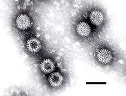
Electron micrograph of rotaviruses. The bar = 100 nm
Virus classification
Group: Group III (dsRNA)
Families

Birnaviridae
Cystoviridae
Hypoviridae
Partitiviridae
Reoviridae
Totiviridae

Double-stranded (ds) RNA viruses are a diverse group of viruses that vary widely in host range (humans, animals, plants, fungi, and bacteria), genome segment number (one to twelve), and virion organization (T-number, capsid layers, or turrets). Members of this group include the rotaviruses, known globally as a common cause of gastroenteritis in young children, and bluetongue virus, an economically important pathogen of cattle and sheep.

Viruses with dsRNA genomes are currently grouped into six families: Reoviridae, Birnaviridae, Totiviridae, Partitiviridae, Hypoviridae, and Cystoviridae. Of these six families, the Reoviridae is the largest and most diverse in terms of host range.

In recent years virus particle assembly, virus-cell interactions, and viral pathogenesis, approaches for the development of novel antiviral strategies and/or agents can be designed.


Reoviridae

Reoviridae are currently classified into nine genera. The genomes of these viruses consist of 10 to 12 segments of dsRNA, each generally encoding one protein. The mature virions are non-enveloped. Their capsids, formed by multiple proteins, have icosahedral symmetry and are arranged generally in concentric layers. A distinguishing feature of the dsRNA viruses, irrespective of their family association, is their ability to carry out transcription of the dsRNA segments, under appropriate conditions, within the capsid. In all these viruses, the enzymes required for endogenous transcription are thus part of the virion structure.


Orthoreoviruses

The orthoreoviruses (reoviruses) are the prototypic members of the virus Reoviridae family and representative of the turreted members, which comprise about half the genera. Like other members of the family, the reoviruses are non-enveloped and characterized by concentric capsid shells that encapsidate a segmented dsRNA genome. In particular, reovirus has eight structural proteins and ten segments of dsRNA. A series of uncoating steps and conformational changes accompany cell entry and replication. High-resolution structures are known for almost all of the proteins of mammalian reovirus (MRV), which is the best-studied genotype. Electron cryo-microscopy (cryoEM) and X-ray crystallography have provided a wealth of structural information about two specific MRV strains, type 1 Lang (T1L) and type 3 Dearing (T3D).


Cypovirus

The cytoplasmic polyhedrosis viruses (CPVs) form the genus Cypovirus of the family Reoviridae. CPVs are classified into 14 species based on the electrophoretic migration profiles of their genome segments. Cypovirus has only a single capsid shell, which is similar to the orthoreovirus inner core. CPV exhibits striking capsid stability and is fully capable of endogenous RNA transcription and processing. The overall folds of CPV proteins are similar to those of other reoviruses. However, CPV proteins have insertional domains and unique structures that contribute to their extensive intermolecular interactions. The CPV turret protein contains two methylase domains with a highly conserved helix-pair/β-sheet/helix-pair sandwich fold but lacks the β-barrel flap present in orthoreovirus λ2. The stacking of turret protein functional domains and the presence of constrictions and A spikes along the mRNA release pathway indicate a mechanism that uses pores and channels to regulate the highly coordinated steps of RNA transcription, processing, and release.


Rotaviruse

Rotavirus is the most common cause of acute gastroenteritis in infants and young children worldwide. This virus contains a dsRNA genome and is a member of the Reoviridae family. The genome of rotavirus consists of eleven segments of dsRNA. Each genome segment codes for one protein with the exception of segment 11, which codes for two proteins. Among the twelve proteins, six are structural and six are non-structural proteins.


Bluetongue virus

The members of Orbivirus genus within the Reoviridae family are arthropod-borne viruses and are responsible for high morbidity and mortality in ruminants. Bluetongue virus (BTV) which causes disease in livestock (sheep, goat, cattle) has been in the forefront of molecular studies for the last three decades and now represents the best understood orbivirus at the molecular and structural levels. BTV, like other members of the family, is a complex non-enveloped virus with seven structural proteins and a RNA genome consisting of 10 variously sized dsRNA segments.


Phytoreoviruses

Phytoreoviruses are non-turreted reoviruses that are major agricultural pathogens, particularly in Asia. One member of this family, Rice Dwarf Virus (RDV), has been extensively studied by electron cryomicroscopy and x-ray crystallography. From these analyses, atomic models of the capsid proteins and a plausible model for capsid assembly have been derived. While the structural proteins of RDV share no sequence similarity to other proteins, their folds and the overall capsid structure are similar to those of other Reoviridae.


L-A Virus

The L-A dsRNA virus of the yeast Saccharomyces cerevisiae has a single 4.6 kb genomic segment that encodes its major coat protein, Gag (76 kDa) and a Gag-Pol fusion protein (180 kDa) formed by a -1 ribosomal frameshift. L-A can support the replication and encapsidation in separate viral particles of any of several satellite dsRNAs, called M dsRNAs, each of which encodes a secreted protein toxin (the killer toxin) and immunity to that toxin. L-A and M are transmitted from cell to cell by the cytoplasmic mixing that occurs in the process of mating. Neither is naturally released from the cell or enters cells by other mechanisms, but the high frequency of yeast mating in nature results in the wide distribution of these viruses in natural isolates. Moreover, the structural and functional similarities with dsRNA viruses of mammals has made it useful to consider these entities as viruses.


Infectious bursal disease virus

Infectious bursal disease virus (IBDV) is the best-characterized member of the family Birnaviridae. These viruses have bipartite dsRNA genomes enclosed in single-layered icosahedral capsids with T = 13l geometry. IBDV shares functional strategies and structural features with many other icosahedral dsRNA viruses, except that it lacks the T = 1 (or pseudo T = 2) core common to the Reoviridae, Cystoviridae, and Totiviridae. The IBDV capsid protein exhibits structural domains that show homology to those of the capsid proteins of some positive-sense single-stranded RNA viruses, such as the nodaviruses and tetraviruses, as well as the T = 13 capsid shell protein of the Reoviridae. The T = 13 shell of the IBDV capsid is formed by trimers of VP2, a protein generated by removal of the C-terminal domain from its precursor, pVP2. The trimming of pVP2 is performed on immature particles as part of the maturation process. The other major structural protein, VP3, is a multifunctional component lying under the T = 13 shell that influences the inherent structural polymorphism of pVP2. The virus-encoded RNA-dependent RNA polymerase, VP1, is incorporated into the capsid through its association with VP3. VP3 also interacts extensively with the viral dsRNA genome.

Bacteriophage Φ6

Bacteriophage Φ6, is a member of the Cystoviridae family. It infects Pseudomonas bacteria (typically plant-pathogenic P. syringae). It has a three-part, segmented, double-stranded RNA genome, totalling ~13.5 kb in length. Φ6 and its relatives have a lipid membrane around their nucleocapsid, a rare trait among bacteriophages. It is a lytic phage, though under certain circumstances has been observed to display a delay in lysis which may be described as a "carrier state".

RNA virus

An RNA virus is a virus that has RNA (ribonucleic acid) as its genetic material. This nucleic acid is usually single-stranded RNA (ssRNA) but may be double-stranded RNA (dsRNA). The ICTV classifies RNA viruses as those that belong to Group III, Group IV or Group V of the Baltimore classification system of classifying viruses, and does not consider viruses with DNA intermediates as RNA viruses. Notable human diseases caused by RNA viruses include SARS, influenza and hepatitis C.

Another term for RNA viruses that explicitly excludes retroviruses is ribovirus.


Characteristics

Single-stranded RNA viruses and RNA Sense

RNA viruses can be further classified according to the sense or polarity o

f their RNA into negative-sense and positive-sense, or ambisense RNA viruses. Positive-sense viral RNA is identical to viral mRNA and thus can be immediately translated by the host cell. Negative-sense viral RNA is complementary to mRNA and thus must be converted to positive-sense RNA by an RNA polymerase before translation. As such, purified RNA of a positive-sense virus can directly cause infection though it may be less infectious than the whole virus particle. Purified RNA of a negative-sense virus is not infectious by itself as it needs to be transcribed into positive-sense RNA. Ambisense RNA viruses resemble negative-sense RNA viruses, except they also translate genes from the positive strand.

Double-stranded RNA viruses

The double-stranded (ds)RNA viruses represent a diverse group of viruses that vary widely in host range (humans, animals, plants, fungi, and bacteria), genome segment number (one to twelve), and virion organization (T-number, capsid layers, or turrets). Members of this group include the rotaviruses, renowned globally as the commonest cause of gastroenteritis in young children, and bluetongue virus, an economically important pathogen of cattle and sheep. In recent years, remarkable progress has been made in determining, at atomic and subnanometeric levels, the structures of a number of key viral proteins and of the virion capsids of several dsRNA viruses, highlighting the significant parallels in the structure and replicative processes of many of these viruses.

Mutation rates

RNA viruses generally have very high mutation rates as they lack DNA polymerases which can find and fix mistakes, and are therefore unable to conduct DNA repair of damaged genetic material. DNA viruses have considerably lower mutation rates due to the proof-reading ability of DNA polymerases within the host cell. Retroviruses have a high mutation rate even though their DNA intermediate integrates into the host genome (and is thus subject to host DNA proofreading once integrated), because errors during reverse transcription are embedded into both strands of DNA prior to integration.

Although RNA usually mutates rapidly, recent work found that the SARS virus and related RNA viruses contain a gene that mutates very slowly. The gene in question has a complex three-dimensional structure which is hypothesized to provide a chemical function necessary for viral propagation, perhaps as a ribozyme. If so, most mutations would render it unfit for that purpose and would not propagate.


Replication

Animal RNA viruses are classified into three distinct groups depending on their genome and mode of replication (and the numerical groups based on the older Baltimore classification):

  • Double-stranded RNA viruses (Group III) contain from one to a dozen different RNA molecules, each of which codes for one or more viral proteins.
  • Positive-sense ssRNA viruses (Group IV) have their genome directly utilized as if it were mRNA, producing a single protein which is modified by host and viral proteins to form the various proteins needed for replication. One of these includes RNA-d ependent RNA polymerase, which copies the viral RNA to form a double-stranded replicative form, in turn this directs the formation of new virions.
  • Negative-sense ssRNA viruses (Group V) must have their genome copied by an RNA polymerase to form positive-sense RNA. This means that the virus must bring along with it the RNA-dependent RNA polymerase enzyme. The positive-sense RNA molecule then acts as viral mRNA, which is translated into proteins by the host ribosomes. The resultant protein goes on to direct the synthesis of new virions, such as capsid proteins and RNA replicase, which is used to produce new negative-sense RNA molecules.

Retroviruses (Group VI) have a single-stranded RNA genome but are generally not considered RNA viruses because they use DNA intermediates to replicate. Reverse transcriptase, a viral enzyme that comes from the virus itself after it is uncoated, converts the viral RNA into a complementary strand of DNA, which is copied to produce a double stranded molecule of viral DNA. After this DNA is integrated, expression of the encoded genes may lead the formation of new virions.


Group III - dsRNA viruses

  • Family Birnaviridae
  • Family Chrysoviridae
  • Family Cystoviridae
  • Family Hypoviridae
  • Family Partitiviridae
  • Family Reoviridae - includes Rotavirus
  • Family Totiviridae
  • Unassigned genera

    • Endornavirus

Group IV - positive-sense ssRNA viruses

  • Order Nidovirales
    • Family Arteriviridae
    • Family Coronaviridae - includes Coronavirus, SARS
    • Family Roniviridae
  • Unassigned
    • Family Astroviridae
    • Family Barnaviridae
    • Family Bromoviridae
    • Family Caliciviridae - includes Norwalk virus
    • Family Closteroviridae
    • Family Comoviridae
    • Family Dicistroviridae
    • Family Flaviviridae - includes Yellow fever virus, West Nile virus, Hepatitis C virus, Dengue fever virus
    • Family Flexiviridae
    • Family Leviviridae
    • Family Luteoviridae - includes Barley yellow dwarf virus
    • Family Marnaviridae
    • Family Narnaviridae
    • Family Nodaviridae
    • Family Picornaviridae - includes Poliovirus, the common cold virus, Hepatitis A virus
    • Family Potyviridae
    • Family Sequiviridae
    • Family Tetraviridae
    • Family Togaviridae - includes Rubella virus, Ross River virus, Sindbis virus,Chikungunya virus
    • Family Tombusviridae
    • Family Tymoviridae
    • Unassigned genera
      • Genus Benyvirus
      • Genus Cheravirus
      • Genus Furovirus
      • Genus Hepevirus - includes Hepatitis E virus
      • Genus Hordeivirus
      • Genus Idaeovirus
      • Genus Ourmiavirus
      • Genus Pecluvirus
      • Genus Pomovirus
      • Genus Sadwavirus
      • Genus Sobemovirus
      • Genus Tobamovirus - includes tobacco mosaic virus
      • Genus Tobravirus
      • Genus Umbravirus


Group V - negative-sense ssRNA viruses

  • Order Mononegavirales
    • Family Bornaviridae - Borna disease virus
    • Family Filoviridae - includes Ebola virus, Marburg virus
    • Family Paramyxoviridae - includes Measles virus, Mumps virus, Nipah virus, Hendra virus
    • Family Rhabdoviridae - includes Rabies virus
  • Unassigned
    • Family Arenaviridae - includes Lassa virus
    • Family Bunyaviridae - includes Hantavirus
    • Family Orthomyxoviridae - includes Influenza viruses
    • Unassigned genera:
      • Genus Deltavirus
      • Genus Ophiovirus
      • Genus Tenuivirus
      • Genus Varicosavirus


Antisense RNA

Antisense RNA is single-stranded RNA that is complementary to a messenger RNA (mRNA) strand transcribed within a cell. Antisense RNA may be introduced into a cell to inhibit translation of a complementary mRNA by base pairing to it and physically obstructing the translation machinery. This effect is therefore stoichiometric. An example of naturally occurring mRNA antisense mechanism is the hok/sok system of the E.coli R1 plasmid. Antisense RNA has long been thought of as a promising technique for disease therapy; the only such case to have reached the market is the drug fomivirsen. One commentator has characterized antisense RNA as one of "dozens of technologies that are gorgeous in concept, but exasperating in [commercialization]". Generally, antisense RNA still lack effective design, biological activity, and efficient route of administration.

Historically, the effects of antisense RNA have often been confused with the effects of RNA interference, a related process in which double-stranded RNA fragments called small interfering RNAs trigger catalytically mediated gene silencing, most typically by targeting the RNA-induced silencing complex (RISC) to bind to and degrade the mRNA. Attempts to genetically engineer transgenic plants to express antisense RNA instead activate the RNAi pathway, although the processes result in differing magnitudes of the same downstream effect, gene silencing. Well-known examples include the Flavr Savr tomato and two cultivars of ringspot-resistant papaya.

Transcription of longer cis-antisense transcripts is a common phenomenon in the mammalian transcriptome. Although the function of some cases have been described, such as the Zeb2/Sip1 antisense RNA, no general function has been elucidated. In the case of Zeb2/Sip1, the antisense noncoding RNA is opposite the 5' splice site of an intron in the 5'UTR of the Zeb2 mRNA. Expression of the antisense ncRNA prevents splicing of an intron that contains a ribosome entry site necessary for efficient expression of the Zeb2 protein. Transcription of long antisense ncRNAs is often concordant with the associated protein-coding gene, but more detailed studies have revealed that the relative expression patterns of the mRNA and antisense ncRNA are complex.

Sense (molecular biology)

Sense, when applied in a molecular biology context, is a general concept used to compare the polarity of nucleic acid molecules, such as DNA or RNA, to other nucleic acid molecules. Depending on the context within molecular biology, sense may have slightly different meanings.


DNA sense

Molecular biologists call a DNA single strand or sequence sense (or positive sense) if an RNA version of the same sequence is translated or translatable into protein, and they call its complement antisense (or negative sense). Sometimes the phrase coding strand is encountered; however, protein coding and non-coding RNA's can be transcribed similarly from both strands, in some cases being transcribed in both directions from a common promoter region, or being transcribed from within introns, on both strands.

Antisense DNA

Schematic showing how antisense DNA strands can interfere with protein translation.

Antisense molecules interact with complementary strands of nucleic acids, modifying expression of genes.

Some regions within a double strand of DNA code for genes, which are usually instructions specifying the order of amino acids in a protein along with regulatory sequences, splicing sites, noncoding introns and other complicating details. For a cell to use this information, one strand of the DNA serves as a template for the synthesis of a complementary strand of RNA. The template DNA strand is called the transcribed strand with antisense sequence and the mRNA transcript is said to be sense sequence (the complement of antisense). Because the DNA is double-stranded, the strand complementary to the antisense sequence is called non-transcribed strand and has the same sense sequence as the mRNA transcript (though T bases in DNA are substituted with U bases in RNA).

DNA strand 1: sense strand

DNA strand 2: antisense strand (copied to)→ RNA strand (sense)

Many forms of antisense have been developed and can be broadly categorized into enzyme-dependent antisense or steric blocking antisense.

Enzyme-dependent antisense includes forms dependent on RNase H activity to degrade target mRNA, including single-stranded DNA, RNA, and phosphorothioate antisense. The R1 plasmid hok/sok system is an example of mRNA antisense regulation process, through enzymatic degradation of the resulting RNA duplex. Double stranded RNA acts as enzyme-dependent antisense through the RNAi/siRNA pathway, involving target mRNA recognition through sense-antisense strand pairing followed by target mRNA degradation by the RNA-induced silencing complex (RISC).

Steric blocking antisense (RNase-H independent antisense) interferes with gene expression or other mRNA-dependent cellular processes by binding to a target sequence of mRNA and getting in the way of other processes. Steric blocking antisense includes 2'-O alkyl (usually in chimeras with RNase-H dependent antisense), peptide nucleic acid (PNA), locked nucleic acid (LNA) and Morpholino antisense.

Antisense nucleic acid molecules have been used experimentally to bind to mRNA and prevent expression of specific genes. Antisense therapies are also in development; in the USA, the Food and Drug Administration (FDA) has approved a phosphorothioate antisense oligo, fomivirsen (Vitravene), for human therapeutic use.

Cells can produce antisense RNA molecules naturally, which interact with complementary mRNA molecules and inhibit their expression.


Ambisense

A single-stranded genome which contains both positive-sense and negative-sense is said to be ambisense. Bunya viruses have 3 single-stranded RNA (ssRNA) fragments containing both positive-sense and negative-sense sections; arenaviruses are also ssRNA viruses with an ambisense genome, as they have 2 fragments which are mainly negative-sense except for part of the 5' ends of the large and small segments of their genome.


Antisense mRNA

Antisense mRNA is an mRNA transcript that is complementary to endogenous mRNA. In other words, it is a non-coding strand complementary to the coding sequence of mRNA; this is similar to negative-sense viral RNA. Introducing a transgene coding for antisense mRNA is a technique used to block expression of a gene of interest. Radioactively-labelled antisense mRNA can be used to show the level of transcription of genes in various cell types. Some alternative antisense structural types are being experimentally applied as antisense therapy, with at least one antisense therapy approved for use in humans.


RNA sense

In virology, the genome of a RNA virus can be said to be either positive-sense, also known as a "plus-strand", or negative-sense, also known as a "minus-strand". In most cases, the terms sense and strand are used interchangeably, making such terms as positive-strand equivalent to positive-sense, and plus-strand equivalent to plus-sense. Whether a virus is positive-sense or negative-sense can be used as a basis for classifiying viruses.

Positive-sense

Positive-sense (5' to 3') viral RNA signifies that a particular viral RNA sequence may be directly translated into the desired viral proteins. Therefore, in positive-sense RNA viruses, the viral RNA genome can be considered viral mRNA, and can be immediately translated by the host cell. Unlike negative-sense RNA, positive-sense RNA is of the same sense as mRNA. Some viruses (e.g. Coronaviridae) have positive-sense genomes which can act as mRNA and be used directly to synthesise proteins without the help of a complementary RNA intermediate. Because of this, these viruses do not need to have an RNA transcriptase packaged into the virion.

Negative-sense

Negative-sense (3' to 5') viral RNA is complementary to the viral mRNA and thus must be converted to positive-sense RNA by an RNA polymerase prior to translation. Negative-sense RNA (like DNA) has a nucleotide sequence complementary to the mRNA that it encodes. Like DNA, this RNA cannot be translated into protein directly. Instead, it must first be transcribed into a positive-sense RNA which acts as an mRNA. Some viruses (Influenza, for example) have negative-sense genomes and so must carry an RNA polymerase inside the virion.


Magnetic immunoassay

Magnetic immunoassay (MIA) is a novel type of diagnostic immunoassay utilizing magnetic beads as labels in lieu of conventional enzymes (ELISA), radioisotopes (RIA) or fluorescent moieties (fluorescent immunoassays). This assay involves the specific binding of an antibody to its antigen, where a magnetic label is conjugated to one element of the pair. The presence of magnetic beads is then detected by a magnetic reader (magnetometer) which measures the magnetic field change induced by the beads. The signal measured by the magnetometer is proportional to the analyte (virus, toxin, bacteria, cardiac marker,etc.) quantity in the initial sample.


Magnetic labels

Magnetic beads are comprised of nanometric-sized iron oxide particles encapsulated or glued together with polymers. These magnetic beads range from 35nm up to 4.5μm. The component magnetic nanoparticles range from 5 to 50nm and exhibit a unique quality referred to as superparamagnetism in the presence of an externally applied magnetic field. First discovered by Frenchman Louis Néel, Nobel Physics Prize winner in 1970, this superparamagnetic quality has already been utilized for medical application in Magnetic Resonance Imaging (MRI) and in biological separations, but not yet for labeling in commercial diagnostic applications. Magnetic labels exhibit several features very well adapted for such applications:

  • they are not affected by reagent chemistry or photo-bleaching and are therefore stable over time,
  • the magnetic background in a biomolecular sample is usually insignificant,
  • sample turbidity or staining have no impact on magnetic properties,
  • magnetic beads can be manipulated remotely by magnetism.

Magnetometers

A simple instrument can detect the presence and measure the total magnetic signal of a sample, however the challenge of developing an effective MIA is to separate naturally occurring magnetic background (noise) from the weak magnetically labeled target (signal). Various approaches and devices have been employed to achieve a meaningful signal-to-noise ratio (SNR) for bio-sensing applications: giant magneto-resistive sensors and spin valves, piezo-resistive cantilevers, inductive sensors, superconducting quantum interference devices, anisotropic magneto-resistive rings, and miniature Hall sensors. But improving SNR often requires a complex instrument to provide repeated scanning and extrapolation through data processing, or precise alignment of target and sensor of miniature and matching size. Beyond this requirement, MIA that exploits the non-linear magnetic properties of magnetic labels can effectively utilize the intrinsic ability of a magnetic field to pass through plastic, water, nitrocellulose, and other materials, thus allowing for true volumetric measurements in various immunoassay formats. Unlike conventional methods that measure the susceptibility of superparamagnetic materials, a MIA based on non-linear magnetization eliminates the impact of linear dia- or paramagnetic materials such as sample matrix, consumable plastics and/or nitrocellulose. Although the intrinsic magnetism of these materials is very weak, with typical susceptibility values of –10-5 (dia) or +10-3 (para), when one is investigating very small quantities of superparamagnetic materials, such as nanograms per test, the background signal generated by ancillary materials cannot be ignored. In MIA based on non-linear magnetic properties of magnetic labels the beads are exposed to an alternating magnetic field at two frequencies, f1 and f2. In the presence of non-linear materials such as superparamagnetic labels, a signal can be recorded at combinatorial frequencies, for example, at f = f1 ± 2×f2. This signal is exactly proportional to the amount of magnetic material inside the reading coil.

This technology,makes magnetic immunoassay possible in a variety of formats such as:

  • conventional lateral flow test by replacing gold labels with magnetic labels,
  • vertical flow tests allowing for the interrogation of rare analytes (such as bacteria) in large-volume samples
  • microfluidic applications and biochip

It was also described for in vivo applications and for multiparametric testing.


Secondary antibody

The primary antibody (in purple) binds to an antigen (in red). A labeled secondary antibody (in green), then binds to the primary antibody. The label is then used to indirectly detect the antigen.

A secondary antibody is an antibody that binds to primary antibodies or antibody fragments. They are typically labeled with probes that make them useful for detection, purification or cell sorting applications.

Secondary antibodies may be polyclonal or monoclonal, and are available with specificity for whole Ig molecules or antibody fragments such as the Fc or Fab regions.

Specific secondary antibodies are usually chosen to work in specific laboratory applications. Frequently, any one of several secondary antibodies perform adequately in a particular application. They are selected according to the source of the primary antibody, the class of the primary antibody (e.g., IgG or IgM), and the kind of label which is preferred. Identifying the optimal secondary antibody is normally done through trial and error.


Applications

Secondary antibodies are used in many biochemical assays including:

  • ELISA, including many HIV tests
  • Western blot
  • Immunostaining
  • Immunohistochemistry
  • Immunocytochemistry

Nomenclature of monoclonal antibodies

Complete list of stems for
monoclonal antibody nomenclature
Prefix Target Source Suffix
variable -vi(r)- viral
-u- human -mab
-ba(c)- bacterial
-o- mouse
-li(m)- immune system
-a- rat
-le(s)- infectious lesions -e- hamster
-ci(r)- cardiovascular
-i- primate
-fu(ng)- fungal
-xi- chimeric
-ne(r)- nervous system
-zu- humanized
-ki(n)- interleukin as target -axo- rat/murine hybrid
-mu(l)- musculoskeletal
-xizu- chimeric + humanized
-o(s)- bone
-tox(a)- toxin as target
-anib(i)- angiogenesis
-co(l)- colonic tumor
-me(l)- melanoma
-ma(r)- mammary tumor
-go(t)- testicular tumor
-go(v)- ovarian tumor
-pr(o)- prostate tumor
-tu(m)- miscellaneous tumor

The nomenclature of monoclonal antibodies is a naming scheme for assigning generic, or nonproprietary, names to a group of medicines called monoclonal antibodies. This scheme is used for both the World Health Organization’s International Nonproprietary Names and the United States Adopted Names. In general, suffixes are used to identify a class of medicines; all monoclonal antibody pharmaceuticals end with the suffix -mab. However, different infixes are used depending on the structure and function of the medicine.


Components

Infix for origin/source

The infix preceding the -mab suffix denotes the animal origin of the antibodies. Although the original monoclonal antibodies were produced in mice (infix, -o-), these antibodies are recognized as foreign by human immune systems and may be rapidly cleared, provoke an allergic reaction, or both. Therefore, parts of the antibody may be replaced with human sequences. If the constant region is replaced with the human form, it is termed chimeric and the infix used is -xi-. Part of the variable regions may also be substituted, in which case it is termed humanized and the infix used is -zu-. Antibodies originating in humans use -u-.

Infix for target

The infix preceding the source of the antibodies refers to medicine’s target. Most of these consist of a consonant, vowel, then another consonant. For ease of pronunciation and to avoid awkwardness, the final consonant is dropped if the following infix begins with a consonant (such as -zu- or -xi-). Examples of these include -ci(r)- for the circulatory system and -tu(m)- for miscellaneous tumors (cancers).

Prefix and second word

Finally, the prefix carries no special meaning and should be unique for each medicine. A second word may be added if there is another substance attached or linked.


Examples

Abciximab is a commonly used medication to prevent platelets from clumping together. It can be broken down into ab- + -ci(r)- + -xi- + -mab. Therefore, it is a chimeric monoclonal antibody used on the cardiovascular system.

Another example is the breast cancer medication trastuzumab, which can be broken down into tras- + -tu(m)- + -zu- + -mab. Therefore, it is a humanized monoclonal antibody used against a tumor.

Nanobodies

Nanobodies (aka single domain antibodies or VHH antibodies) are a type of antibodies derived from camelids, and are much smaller than traditional antibodies. Standard antibodies are giants by molecular standards, since each one is a conglomerate of two heavy protein chains and two light chains, intricately folded and garnished with elaborate sugars. Nanobodies, however, are relatively simple proteins about a tenth the size of human antibodies and just a few nanometers in length. After the discovery that camelidae (camels and llamas) possess fully functional antibodies that lack light chains, the nanobody technology was developed to exploit these smaller heavy-chain-only constructs. Nanobodies are being researched for multiple pharmaceutical applications and have potential for use in cancer and Alzheimer's Disease treatments.

Traditional therapeutic monoclonal antibodies (MAbs) must be stored at near freezing temperatures to prevent their destruction. Antibodies are not suited for oral administration because they are digested quickly in the gut, and are not usually useful for treating diseases of the brain because they do not easily permeate the blood-brain barrier. Additionally, therapeutic antibodies are not well suited to target large tumors because they are held to the periphery of solid tumors. Many illnesses are thus unreachable by monoclonals, and patients who use MAb therapies must receive them by injection or infusion at a clinic. For certain conditions in which the traditional MAbs do not work well, and even for some in which they currently do, simpler, smaller proteins like nanobodies might perform better, be easier to make, easier to handle, easier to admister, and be more affordable. (2)

In 1989 a group of biologists led by Raymond Hamers at the Free University (Brussels, Belgium) investigated an odd observation handed in as part of a student project on parasite immunodefense in dromedary camels (the one-humped Arabian variety) and water buffalo. One of the tests for antibodies in the dromedary blood seemed to show an error: in addition to normal four-chain antibodies it indicated the presence of simpler antibodies composed solely of a pair of heavy chains.

After several years of investigation, Hamers and his colleagues published their discovery in Nature in 1993. In dromedaries — and also in two-humped Asian camels and South American llamas — about half the antibodies circulating in the blood lack a light chain. Equally surprising, they found that these "incomplete" antibodies are able to grasp their targets just as firmly as normal antibodies do, with affinities for their targets virtually equal to a full antibody 10 times their size.

These shortened proteins were also more chemically agile, able to engage targets — including the active sites of enzymes and clefts in cell membranes — too small to admit an antibody.

Because nanobodies are so much smaller than antibodies and are not hydrophobes (as are standard human antibodies), they are more resistant to heat and pH, and may retain their activity as they pass through the gastrointestinal tract, raising the prospect of oral nanobody pills to treat inflammatory bowel disease, colon cancer and other disorders of the gut.

Polyclonal antibodies

Polyclonal antibodies (or antisera) are antibodies that are derived from different B cell lines. They are a mixture of immunoglobulin molecules secreted against a specific antigen, each recognising a different epitope.


Production

These antibodies are typically produced by immunization of a suitable mammal, such as a mouse, rabbit or goat. Larger mammals are often preferred as the amount of serum that can be collected is greater. An antigen is injected into the mammal. This induces the B-lymphocytes to produce IgG immunoglobulins specific for the antigen. This polyclonal IgG is polyclonal purified from the mammal’s serum.

By contrast, monoclonal antibodies are derived from a single cell line.

Many methodologies exist for polyclonal antibody production in laboratory animals. Institutional guidelines governing animal use and procedures relating to these methodologies are generally oriented around humane considerations and appropriate conduct for adjuvant (agents which modify the effect of other agents while having few if any direct effects when given by themselves) use. This includes adjuvant selection, routes and sites of administration, injection volumes per site and number of sites per animal. Institutional policies generally include allowable volumes of blood per collection and safety precautions including appropriate restraint and sedation or anesthesia of animals for injury prevention to animals or personnel.

The primary goal of antibody production in laboratory animals is to obtain high titer, high affinity antisera for use in experimentation or diagnostic tests. Adjuvants are used to improve or enhance an immune response to antigens. Most adjuvants provide for an injection site, antigen depot which allows for a slow release of antigen into draining lymph nodes.

Many adjuvants also contain or act directly as:

  1. surfactants which promote concentration of protein antigens molecules over a large surface area, and
  2. immunostimulatory molecules or properties. Adjuvants are generally used with soluble protein antigens to increase antibody titers and induce a prolonged response with accompanying memory.

Such antigens by themselves are generally poor immunogens. Most complex protein antigens induce multiple B-cell clones during the immune response, thus, the response is polyclonal. Immune responses to non-protein antigens are generally poorly or enhanced by adjuvants and there is no system memory.


Animal selection

Animals frequently used for polyclonal antibody production include chickens, goats, guinea pigs, hamsters, horses, mice, rats, and sheep. However, the rabbit is the most commonly used laboratory animal for this purpose. Animal selection should be based upon:

  1. the amount of antibody needed,
  2. the relationship between the donor of the antigen and the recipient antibody producer (generally the more distant the phylogenetic relationship, the greater the potential for high titer antibody response) and
  3. the necessary characteristics [e.g., class, subclass (isotype), complement fixing nature] of the antibodies to be made. Immunization and phlebotomies are stress associated and, at least when using rabbits and rodents, specific pathogen free (SPF) animals are preferred. Use of such animals can dramatically reduce morbidity and mortality due to pathogenic organisms, especially Pasteurella multocida in rabbits.

Goats or horses are generally used when large quantities of antisera are required. Many investigators favor chickens because of their phylogenetic distance from mammals. Chickens transfer high quantities of IgY (IgG) into the egg yolk and harvesting antibodies from eggs eliminates the need for the invasive bleeding procedure. One week’s eggs can contain 10 times more antibodies than the volume of rabbit blood obtained from one weekly bleeding. However, there are some disadvantages when using certain chicken derived antibodies in immunoassays. Chicken IgY does not fix mammalian complement component C1 and it does not perform as a precipitating antibody using standard solutions.

Although mice are used most frequently for monoclonal antibody production, their small size usually prevents their use for sufficient quantities of polyclonal, serum antibodies. However, polyclonal antibodies in mice can be collected from ascites fluid using any one of a number of ascites producing methodologies.

When using rabbits, young adult animals (2.5–3.0 kg or 5.5-6.5lbs) should be used for primary immunization because of the vigorous antibody response. Immune function peaks at puberty and primary responses to new antigens decline with age. Female rabbits are generally preferred because they are more docile and are reported to mount a more vigorous immune response than males. At least two animals per antigen should be used when using outbred animals. This principle reduces potential total failure resulting from non-responsiveness to antigens of individual animals.


Antigen preparation

The size, extent of aggregation and relative nativity of protein antigens can all dramatically affect the quality and quantity of antibody produced. Small polypeptides (<10>

Keyhole limpet hemocyanin (KLH) and bovine serum albumin are two widely used carrier proteins. Poly-L-lysine has also been used successfully as a backbone for peptides. Although the use of Poly-L-lysine reduces or eliminates production of antibodies to foreign proteins, it may result in failure of peptide-induced antibody production. Recently, liposomes have been successfully used for delivery of small peptides and this technique is more efficient than delivery with oily emulsion adjuvants.

Antigen quantity

Selection of antigen quantity for immunization varies with the properties of the antigen and the adjuvant selected. In general, microgram to milligram quantities of protein in adjuvant are necessary to elicit high titer antibodies. Antigen dosage is generally species, rather than body weight, associated. The so called “window” of immunogenicity in each species is broad but too much or too little antigen can induce tolerance, suppression or immune deviation towards cellular immunity rather than a satisfactory humoral response. Optimal and usual protein antigen levels for immunizing specific species have been reported in the following ranges:

  1. rabbit, 50–1000 µg;
  2. mouse, 10–200 µg;
  3. guinea pig, 50–500 µg; and
  4. goat, 250–5000 µg. Optimal “priming” doses are reported to be at the low end of each range.

The affinity of serum antibodies increases with time (months) after injection of antigen-adjuvant mixtures and as antigen in the system decreases. Widely used antigen dosages for “booster” or secondary immunizations are usually one half to equal the priming dosages. Antigens should be free of preparative byproducts and chemicals such as polyacrylamide gel, SDS, urea, endotoxin, particulate matter and extremes of pH.


Peptide Antibodies

When a peptide is being used to generate the antibody, it is extremely important to design the antigens properly. There are several resources that can aid in the design as well as companies that offer this service. Expasy has aggregated a set of public tools under its ProtScale page that require some degree of user knowledge to navigate. For a more simple peptide scoring tool there is a Antigen Profiler tool available that will enable you to score individual peptide sequences based upon a relation epitope mapping database of previous immunogens used to generate antibodies. Finally, as a general rule peptides should follow some basic criteria.

When examining peptides for synthesis and immunization, it is recommended that certain residues and sequences be avoided due to potential synthesis problems. This includes some of the more common characteristics:

• Extremely long repeats of the same amino acid (e.g. RRRR) • Serine (S), Threonine (T), Alanine (A), and Valine (V) doublets • Ending or starting a sequence with a proline (P) • Glutamine (Q) or Asparagine (N) at the n-terminus • Peptides over weighted with hydrophobic residues (e.g. V,A,L,I, etc…)

Reactivity

Investigators should also consider the status of nativity of protein antigens when used as immunogens and reaction with antibodies produced. Antibodies to native proteins react best with native proteins and antibodies to denatured proteins react best with denatured proteins. If elicited antibodies are to be used on membrane blots (proteins subjected to denaturing conditions) then antibodies should be made against denatured proteins. On the other hand, if antibodies are to be used to react with a native protein or block a protein active site, then antibodies should be made against the native protein. Adjuvants can often alter the nativity of the protein. Generally, absorbed protein antigens in a preformed oil-in-water emulsion adjuvant, retain greater native protein structure than those in water-in-oil emulsions.

Asepticity

Antigens should always be prepared using techniques that ensure that they are free of microbial contamination. Most protein antigen preparations can be sterilized by passage through a 0.22u filter. Septic abscesses often occur at inoculation sites of animals when contaminated preparations are used. This can result in failure of immunization against the targeted antigen.


Adjuvants

There are many commercially available immunologic adjuvants. Selection of specific adjuvants or types varies depending upon whether they are to be used for research and antibody production or in vaccine development. Adjuvants for vaccine use only need to produce protective antibodies and good systemic memory while those for antiserum production need to rapidly induce high titer, high avidity antibodies. No single adjuvant is ideal for all purposes and all have advantages and disadvantages. Adjuvant use generally is accompanied by undesirable side effects of varying severity and duration. Research on new adjuvants focuses on substances which have minimal toxicity while retaining maximum immunostimulation. Investigators should always be aware of potential pain and distress associated with adjuvant use in laboratory animals.

The most frequently used adjuvants for antibody production are Freund’s, Alum, the Ribi Adjuvant System and Titermax.

Freund’s adjuvants

There are two basic types of Freund's adjuvants: Freund’s Complete Adjuvant (FCA) and Freund’s Incomplete Adjuvant (FIA). FCA is a water-in-oil emulsion that localizes antigen for release periods up to 6 months. It is formulated with mineral oil, the surfactant mannide monoleate and heat killed Mycobacterium tuberculosis, Mycobacterium butyricum or their extracts (for aggregation of macrophages at the inoculation site). This potent adjuvant stimulates both cell mediated and humoral immunity with preferential induction of antibody against epitopes of denatured proteins. Although FCA has historically been the most widely used adjuvant, it is one of the more toxic agents due to non-metabolizable mineral oil and it induces granulomatous reactions. Its use is limited to laboratory animals and it should be used only with weak antigens. It should not be used more than once in a single animal since multiple FCA inoculations can cause severe systemic reactions and decreased immune responses. Freund’s Incomplete Adjuvant has the same formulation as FCA but does not contain mycobacterium or its components. FIA usually is limited to booster doses of antigen since it normally much less effective than FCA for primary antibody induction. Freund’s adjuvants are normally mixed with equal parts of antigen preparations to form stable emulsions.

Ribi Adjuvant System

Ribi adjuvants are oil-in-water emulsions where antigens are mixed with small volumes of a metabolizable oil (squalene) which are then emulsified with saline containing the surfactant Tween 80. This system also contains refined mycobacterial products (cord factor, cell wall skeleton) as immunostimulants and bacterial monophosphoryl lipid A. Three different species oriented formulations of the adjuvant system are available. These adjuvants interact with membranes of immune cells resulting in cytokine induction, which enhances antigen uptake, processing and presentation. This adjuvant system is much less toxic and less potent than FCA but generally induces satisfactory amounts of high avidity antibodies against protein antigens.

Titermax

Titermax represents a newer generation of adjuvants that are less toxic and contain no biologically derived materials. It is based upon mixtures of surfactant acting, linear, blocks or chains of nonionic copolymers polyoxypropylene (POP) and polyoxyethylene (POE). These copolymers are less toxic than many other surfactant materials and have potent adjuvant properties which favor chemotaxis, complement activation and antibody production. Titermax adjuvant forms a microparticulate water-in-oil emulsion with a copolymer and metabolizable squalene oil. The copolymer is coated with emulsion stabilizing silica particles which allows for incorporation of large amounts of a wide variety of antigenic materials. The adjuvant active copolymer forms hydrophilic surfaces, which activate complement, immune cells and increased expression of class II major histocompatibility molecules on macrophages. Titermax presents antigen in a highly concentrated form to the immune system, which often results in antibody titers comparable to or higher than FCA.

Specol: Specol is a water in oil adjuvant made of purified mineral oil. It has been reported to induce immune response comparable to Freund's adjuvant in rabbit and other research animal while producing fewer histological lesions.


Humanized antibody

Humanized antibodies or chimeric antibodies are a type of monoclonal antibody that have been synthesized using recombinant DNA technology to circumvent the clinical problem of immune response to foreign antigens. The standard procedure of producing monoclonal antibodies yields mouse antibodies. Although murine antibodies are very similar to human ones there are differences, and the human immune system recognizes mouse antibodies as foreign, rapidly removing them from circulation and causing systemic inflammatory effects.

Humanized antibodies are produced by merging the DNA that encodes the binding portion of a monoclonal mouse antibody with human antibody-producing DNA. One then uses mammalian cell cultures to express this DNA and produce these half-mouse and half-human antibodies that are not as immunogenic as the murine variety.


Alternatives

A solution to this problem would be to generate human antibodies directly from humans. However, this is not easy, primarily because it is not clearly ethical to challenge humans with antigen in order to produce antibody. Furthermore, it is not easy to generate human antibodies against human tissues.


Monoclonal antibody therapy

Each antibody binds only one specific antigen.

Monoclonal antibody therapy is the use of monoclonal antibodies (or mAb) to specifically target cells. The main objective is stimulating the patient's immune system to attack the malignant tumor cells and the prevention of tumor growth by blocking specific cell receptors. Variations exist within this treatment, e.g. radioimmunotherapy, where a radioactive dose localizes on target cell line, delivering lethal chemical doses to the target.


Structure and function of human and therapeutic antibodies

Immunoglobulin G (IgG) antibodies are large heterodimeric molecules, approximately 150 kDa and are composed of two different kinds of polypeptide chain, called the heavy (~50kDa) and the light chain (~25kDa). There are two types of light chains, kappa (κ) and lambda (λ). By cleavage with enzyme papain, the Fab (fragment-antigen binding) part can be separated from the Fc (fragment crystalline) part of the molecule (see image). The Fab fragments contain the variable domains, which consist of three hypervariable amino acid domains responsible for the antibody specificity embedded into constant regions. There are four known IgG subclasses all of which are involved in Antibody-dependent cellular cytotoxicity.

The immune system responds to the environmental factors it encounters on the basis of discrimination between self and non-self. Tumor cells are not specifically targeted by one's immune system since tumor cells are the patient's own cells. Tumor cells, however are highly abnormal, and many display unusual antigens that are either inappropriate for the cell type, its environment, or are only normally present during the organisms' development (e.g. fetal antigens).

Other tumor cells display cell surface receptors that are rare or absent on the surfaces of healthy cells, and which are responsible for activating cellular signal transduction pathways that cause the unregulated growth and division of the tumor cell. Examples include ErbB2, a constitutively active cell surface receptor that is produced at abnormally high levels on the surface of approximately 30% of breast cancer tumor cells. Such breast cancer is known a HER2 positive breast cancer.

Antibodies are a key component of the adaptive immune response, playing a central role in both in the recognition of foreign antigens and the stimulation of an immune response to them. The advent of monoclonal antibody technology has made it possible to raise antibodies against specific antigens presented on the surfaces of tumors.


Origins of monoclonal antibody therapy

Monoclonal antibodies for cancer. ADEPT, antibody directed enzyme prodrug therapy; ADCC, antibody dependent cell-mediated cytotoxicity; CDC, complement dependent cytotoxicity; MAb, monoclonal antibody; scFv, single-chain Fv fragment.

Immunotherapy developed as a technique with the discovery of the structure of antibodies and the development of hybridoma technology, which provided the first reliable source of monoclonal antibodies. These advances allowed for the specific targeting of tumors both in vitro and in vivo. Initial research on malignant neoplasms found MAb therapy of limited and generally short-lived success with malignancies of the blood. Furthermore treatment had to be specifically tailored to each individual patient, thus proving to be impracticable for the routine clinical setting.

Throughout the progression of monoclonal drug development there have been four major antibody types developed: murine, chimeric, humanised and human. Initial therapeutic antibodies were simple murine analogues, which contributed to the early lack of success. It has since been shown that these antibodies have: a short half-life in vivo (due to immune complex formation), limited penetration into tumour sites, and that they inadequately recruit host effector functions. To overcome these difficulties the technical issues initially experienced had to be surpassed. Chimeric and humanized antibodies have generally replaced murine antibodies in modern therapeutic antibody applications. Hybridoma technology has been replaced by recombinant DNA technology, transgenic mice and phage display. Understanding of proteomics has proven essential in identifying novel tumour targets.

Murine monoclonal antibodies

Initially, murine antibodies were obtained by hybridoma technology, for which Kohler and Milstein received a Nobel prize. However the dissimilarity between murine and human immune systems led to the clinical failure of these antibodies, except in some specific circumstances. Major problems associated with murine antibodies included reduced stimulation of cytotoxicity and the formation complexes after repeated administration, which resulted in mild allergic reactions and sometimes anaphylactic shock.

Chimeric and humanized monoclonal antibodies

To reduce murine antibody immunogenicity, murine molecules were engineered to remove immunogenic content and to increase their immunologic efficiency. This was initially achieved by the production of chimeric and humanized antibodies. Chimeric antibodies are composed of murine variable regions fused onto human constant regions. Human gene sequences, taken from the kappa light chain and the IgG1 heavy chain, results in antibodies that are approximately 65% human. This reduces immunogenicity, and thus increases serum half-life.

Humanised antibodies are produced by grafting murine hypervariable amino acid domains into human antibodies. This results in a molecule of approximately 95% human origin. However it has been shown in several studies that humanised antibodies bind antigen much more weakly than the parent murine monoclonal antibody, with reported decreases in affinity of up to several hundredfold. Increases in antibody-antigen binding strength have been achieved by introducing mutations into the complementarity determining regions (CDR), using techniques such as chain-shuffling, randomization of complementarity determining regions and generation of antibody libraries with mutations within the variable regions by error-prone PCR, E-coli mutator strains, and site-specific mutagenesis.

Human monoclonal antibodies

Human monoclonal antibodies are produced using transgenic mice or phage display libraries. Human monoclonal antibodies are produced by transferring human immunoglobulin genes into the murine genome, after which the transgenic mouse is vaccinated against the desired antigen, leading to the production of monoclonal antibodies. Phage display libraries allow the transformation of murine antibodies in vitro into fully human antibodies.


FDA approved therapeutic antibodies

The first FDA-approved therapeutic monoclonal antibody was a murine IgG2a CD3 specific transplant rejection drug, Muromonab (OKT-3), in 1986. This drug found use in solid organ transplant recipients who became steroid resistant. Currently, twenty-one FDA-approved therapies exist, and hundreds of therapies are undergoing clinical trials. Most are concerned with immunological and oncological targets.


FDA approved monoclonal antibodies


Antibody Brand name Approval date Type Target Approved treatment(s) Abciximab
ReoPro 1994 chimeric inhibition of glycoprotein IIb/IIIa Cardiovascular disease
Adalimumab
Humira 2002 human inhibition of TNF-a signalling Inflammatory diseases (mostly auto-immune disorders) Alemtuzumab
Campath 2001 humanized CD52
Chronic lymphocytic leukemia
Basiliximab
Simulect 1998 chimeric IL-2 receptor a Transplant rejection
Bevacizumab
Avastin 2004 humanized vascular endothelial growth factor
Colorectal cancer
Cetuximab
Erbitux 2004 chimeric epidermal growth factor receptor
Colorectal cancer Daclizumab
Zenapax 1997 humanized IL-2 receptor a Transplant rejection Eculizumab
Soliris 2007 humanized complement system protein C5 Inflammatory diseases including paroxysmal nocturnal hemoglobinuria Efalizumab
Raptiva 2002 humanized CD11a
Inflammatory diseases (psoriasis) Ibritumomab tiuxetan
Zevalin 2002 murine CD20
Non-Hodgkin lymphoma (with yttrium-90 or indium-111) Infliximab
Remicade 1998 chimeric inhibition of TNF-a signalling Inflammatory diseases (mostly auto-immune disorders) Muromonab-CD3
Orthoclone OKT3 1986 murine T cell CD3 Receptor Transplant rejection Natalizumab
Tysabri 2006 humanized T cell VLA4 receptor Inflammatory diseases (mainly autoimmune-related multiple sclerosis therapy) Omalizumab
Xolair 2004 humanized immunoglobulin E (IgE) Inflammatory diseases (mainly allergy-related asthma therapy) Palivizumab
Synagis 1998 humanized an epitope of the F protein of RSV Viral infection (especially Respiratory Syncytial Virus (RSV) Panitumumab
Vectibix 2006 human epidermal growth factor receptor Colorectal cancer Ranibizumab
Lucentis 2006 humanized vascular endothelial growth factor
Macular degeneration
Gemtuzumab ozogamicin
Mylotarg 2000 humanized CD33
Acute myelogenous leukemia (with calicheamicin) Rituximab
Rituxan, Mabthera 1997 chimeric CD20
Non-Hodgkin lymphoma
Tositumomab
Bexxar 2003 murine CD20
Non-Hodgkin lymphoma Trastuzumab
Herceptin 1998 humanized ErbB2
Breast cancer
Radioimmunotherapy

Radioimmunotherapy involves the use of radioactively conjugated murine antibodies against cellular antigens. Most research currently involved their application to lymphomas, as these are highly radio-sensitive malignancies. To limit radiation exposure, murine antibodies were especially chosen, as their high immunogenicity promotes rapid clearance from the body. Tositumomab is an exemplar used for non-Hodgkins lymphoma.


Antibody-directed enzyme prodrug therapy (ADEPT)

ADEPT involves the application of cancer associated monoclonal antibodies which are linked to a drug-activating enzyme. Subsequent systemic administration of a non-toxic agent results in its conversion to a toxic drug, and resulting in a cytotoxic effect which can be targeted at malignant cells. The clinical success of ADEPT treatments has been limited to date. However it holds great promise, and recent reports suggest that it will have a role in future oncological treatment.


Drug and gene therapy: Immuno-liposomes

Immunoliposomes are antibody-conjugated liposomes. Liposomes can carry drugs or therapeutic nucleotides and when conjugated with monoclonal antibodies, may be directed against malignant cells. Although this technique is still in its infancy, significant advances have been made. Immunoliposomes have been successfully used in vivo to achieve targeted delivery of tumour-suppressing genes into tumours, using an antibody fragment against the human transferrin receptor. Tissue-specific gene delivery using immunoliposomes has also been achieved in brain, and breast cancer tissue.


Therapeutic Monoclonal Antibody Market Future

Since 2000, the therapeutic market for monoclonal antibodies has grown exponentially. The current “big 5” therapeutic antibodies on the market: Avastin, Herceptin (both oncology), Humira, Remicade (both Autoimmune and Infectious Disease ‘AIID’) and Rituxan (oncology and AIID) accounted for 80% of revenues in 2006.

In the immediate future, it is likely that Genentech/Roche will retain their control over the market (due to ownership of 3 of the “big 5” products), oncology and AIID will remain the mAb segment therapeutic focus (because these are the disease areas addressed by the big 5) and the three most commercially important ‘targets’ for the mAb class will be VEGF (Avastin), TNF-alpha (Remicade and Humira) and CD20 (Rituxan). Experts forecast that the therapeutic antibody market will continue to be dominated by Oncology and AIID segments (82-84 percent) from 2004 to 2011. Furthermore, experts note a potential for change in the balance between Oncology and AIID in the coming years. While Oncology therapeutics dominated the market in 2004, AIID is expected to dominate by 2011.

Monoclonal antibodies

A general representation of the methods used to produce monoclonal antibodies.

Monoclonal antibodies (mAb or moAb) are monospecific antibodies that are identical because they are produced by one type of immune cell that are all clones of a single parent cell. Given (almost) any substance, it is possible to create monoclonal antibodies that specifically bind to that substance; they can then serve to detect or purify that substance. This has become an important tool in biochemistry, molecular biology and medicine. When used as medications, the generic name ends in -mab (see "Nomenclature of monoclonal antibodies").


Discovery

The idea of a "magic bullet" was first proposed by Paul Ehrlich who at the beginning of the 20th century postulated that if a compound could be made that selectively targeted a disease-causing organism, then a toxin for that organism could be delivered along with the agent of selectivity.

In the 1970s the B-cell cancer multiple myeloma was known, and it was understood that these cancerous B-cells all produce a single type of antibody (a paraprotein). This was used to study the structure of antibodies, but it was not yet possible to produce identical antibodies specific to a given antigen.

A process of producing monoclonal antibodies involving human-mouse hybrid cells was described by Jerrold Schwaber in 1973 and remains widely cited among those using human-derived hybridomas, but claims to priority have been controversial. A science history paper on the subject gave some credit to Schwaber for inventing a technique that was widely cited, but stopped short of suggesting that he had been cheated. The invention is generally accredited to Georges Köhler, César Milstein, and Niels Kaj Jerne in 1975; who shared the Nobel Prize in Physiology or Medicine in 1984 for the discovery. The key idea was to use a line of myeloma cells that had lost their ability to secrete antibodies, come up with a technique to fuse these cells with healthy antibody producing B-cells, and be able to select for the successfully fused cells.

In 1988 Greg Winter and his team pioneered the techniques to humanize monoclonal antibodies, removing the reactions that many monoclonal antibodies caused in some patients.


Production

Researchers looking at slides of cultures of cells that make monoclonal antibodies. These are grown in a lab and the researchers are analyzing the products to select the most promising of them.

Monoclonal antibodies can be grown in unlimited quantities in the bottles shown in this picture.

Technician hand-filling wells with a liquid for a research test. This test involves preparation of cultures in which hybrids are grown in large quantities to produce desired antibody. This is effected by fusing myeloma cell and mouse lymphocyte to form a hybrid cell (hybridoma).

Lab technician bathing prepared slides in a solution. This technician prepares slides of monoclonal antibodies for researchers. The cells shown are labeling human breast cancer.

Hybridoma Cell Production

Monoclonal antibodies are typically made by fusing myeloma cells with the spleen cells from a mouse that has been immunized with the desired antigen. However, recent advances have allowed the use of rabbit B-cells. Polyethylene glycol is used to fuse adjacent plasma membranes, but the success rate is low so a selective medium is used in which only fused cells can grow. This is because myeloma cells have lost the ability to synthesize hypoxanthine-guanine-phosphoribosyl transferase (HGPRT), an enzyme necessary for the salvage synthesis of nucleic acids.

This enzyme enables cells to synthesize purines by the salvage pathway, here using an extracellular source of hypoxanthine as a precursor. Ordinarily, the absence of HGPRT is not a problem for the cell because cells have an already existing biochemical pathway, the de novo pathway that they can use to synthesize purines. However, when cells are exposed to Aminopterin (a folic acid analogue, which inhibits Dihydrofolate reductase, DHFR), they are unable to use the de novo pathway and are now fully dependent on the salvage pathway for survival.

The selective culture medium is called HAT medium because it contains Hypoxanthine, Aminopterin, and Thymidine. This medium is selective for fused (hybridoma) cells, because unfused myeloma cells cannot grow because they lack HGPRT. The unfused normal spleen cells cannot grow indefinitely because of their limited life span. Therefore, only hybridoma cells are able to grow indefinitely because the spleen cell partner supplies HGPRT and the myeloma partner gives it immortality (as it is a cancer cell). The fused hybrid cells are called hybridomas, and since they are derived from cancer cells, are immortal and can be grown indefinitely.

This mixture of cells is then diluted and clones are grown from single parent cells on microtitre wells. The antibodies secreted by the different clones are then tested for their ability to bind to the antigen (for example with a test such as ELISA or Antigen Microarray Assay) or immuno-dot blot, and the most productive and stable clone is then grown in culture medium to a high volume.

The hybridomas are grown indefinitely in asuitable cell culture media, or they are injected in mice (in the peritoneal cavity, the gut), they produce tumors containing an antibody-rich fluid called ascites fluid. The medium must be enriched during selection to further favour hybridoma growth. This can be achieved by the use of a layer of feeder fibrocyte cells or supplement medium such as briclone. Production in cell culture is usually preferred as the ascites technique is painful to the animal and if replacement techniques exist, this method is considered unethical.

Recombinant

The production of recombinant monoclonal antibodies involves technologies, referred to as repertoire cloning or phage display/yeast display. Recombinant antibody engineering involves the use of viruses or yeast to create antibodies, rather than mice. These techniques rely on rapid cloning of immunoglobulin gene segments to create libraries of antibodies with slightly different amino acid sequences from which antibodies with desired specificities can be selected. These techniques can be used to enhance the specificity with which antibodies recognize antigens, their stability in various environmental conditions, their therapeutic efficacy, and their detectability in diagnostic applications. Fermentation chambers have been used to produce these antibodies on a large scale.


Applications

Once monoclonal antibodies for a given substance have been produced, they can be used to detect the presence and quantity of this substance, for instance in a Western blot test (to detect a protein on a membrane) or an immunofluorescence test (to detect a substance in a cell). They are also very useful in immunohistochemistry which detect antigen in fixed tissue sections. Monoclonal antibodies can also be used to purify a substance with techniques called immunoprecipitation and affinity chromatography.

Monoclonal antibodies for cancer treatment

One possible treatment for cancer involves monoclonal antibodies that bind only to cancer cell-specific antigens and induce an immunological response against the target cancer cell. Such mAb could also be modified for delivery of a toxin, radioisotope, cytokine or other active conjugate; it is also possible to design bispecific antibodies that can bind with their Fab regions both to target antigen and to a conjugate or effector cell. In fact, every intact antibody can bind to cell receptors or other proteins with its Fc region.

Monoclonal antibodies for cancer. ADEPT, antibody directed enzyme prodrug therapy; ADCC, antibody dependent cell-mediated cytotoxicity; CDC, complement dependent cytotoxicity; MAb, monoclonal antibody; scFv, single-chain Fv fragment.

The illustration below shows all these possibilities:

Chimeric and humanized antibodies

One problem in medical applications is that the standard procedure of producing monoclonal antibodies yields mouse antibodies. Although murine antibodies are very similar to human ones there are differences. The human immune system hence recognizes mouse antibodies as foreign, rapidly removing them from circulation and causing systemic inflammatory effects.

A solution to this problem would be to generate human antibodies directly from humans. However, this is not easy, primarily because it is generally not seen as ethical to challenge humans with antigen in order to produce antibody; the ethics of doing the same to non-humans is a matter of debate. Furthermore, it is not easy to generate human antibodies against human tissues.

Various approaches using recombinant DNA technology to overcome this problem have been tried since the late 1980s. In one approach, one takes the DNA that encodes the binding portion of monoclonal mouse antibodies and merges it with human antibody producing DNA. One then uses mammalian cell cultures to express this DNA and produce these half-mouse and half-human antibodies. (Bacteria cannot be used for this purpose, since they cannot produce this kind of glycoprotein.) Depending on how big a part of the mouse antibody is used, one talks about chimeric antibodies or humanized antibodies. Another approach involves mice genetically engineered to produce more human-like antibodies. Monoclonal antibodies have been generated and approved to treat: cancer, cardiovascular disease, inflammatory diseases, macular degeneration, transplant rejection, multiple sclerosis, and viral infection (see monoclonal antibody therapy).

In August 2006 the Pharmaceutical Research and Manufacturers of America reported that U.S. companies had 160 different monoclonal antibodies in clinical trials or awaiting approval by the Food and Drug Administration.


Examples

Monoclonal antibodies
Type Application Mechanism Mode
infliximab
  • rheumatoid arthritis
  • Crohn's disease
inhibits TNF-α chimeric
basiliximab
  • Acute rejection of kidney transplants
inhibits IL-2 on activated T cells chimeric
abciximab
  • Prevent coagulation in coronary angioplasty
inhibits the receptor GpIIb/IIIa on platelets chimeric
daclizumab
  • Acute rejection of kidney transplants
inhibits IL-2 on activated T cells humanized
gemtuzumab
  • relapsed acute myeloid leukaemia
targets an antigen on leukemia cells humanized
alemtuzumab
  • B cell leukemia
targets an antigen CD52 on T- and B-lymphocytes humanized
rituximab
  • non-Hodgkin's lymphoma
targets phosphoprotein CD20 on B lymphocytes chimeric
palivizumab
  • RSV infections in children
inhibits an RSV protein humanized
trastuzumab
  • anti-cancer therapy for a specific kind of breast cancer
targets the HER2/neu (erbB2) receptor humanized
etanercept
  • rheumatoid arthritis
contains TNF receptor fusion protein
adalimumab
  • rheumatoid arthritis
inhibits TNF-α humanized
Powered By Blogger